Tongue Anatomy, muscles, neurovasculature and histology Kenhub
Anatomy. Under normal circumstances, the tongue is a pink, muscular organ located within the oral cavity proper. It is kept moist by the products of the major and minor salivary glands, which aids the organ as it facilitates deglutition, speech, and gustatory perception.While there is significant variability in the length of the tongue among individuals, on average, the organ is roughly 10 cm.
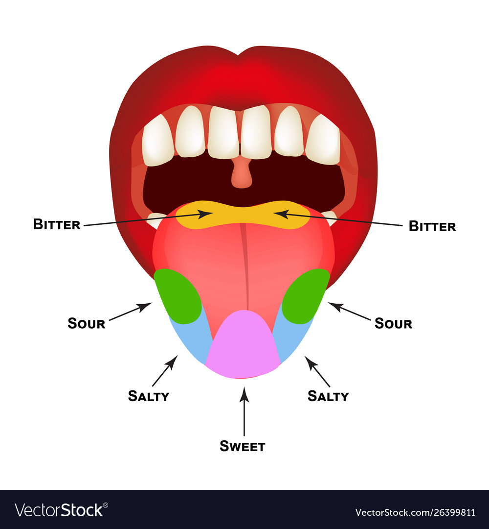
Anatomical structure tongue taste buds on Vector Image
Your tongue is mostly made of muscles. It's anchored inside of your mouth by webs of strong tissue and it's covered by mucosa (a moist, pink lining that covers certain organs and body cavities). Your tongue is also covered with different types of papillae (bumps) and taste buds. You have four different types of taste buds, including:

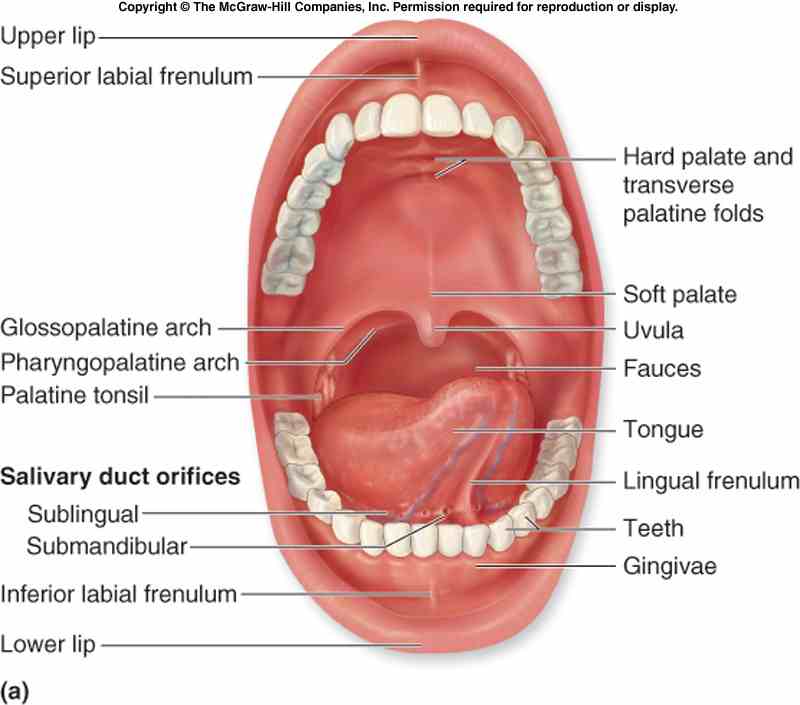
23.3 The Mouth, Pharynx, and Esophagus Anatomy & Physiology
The human tongue is a muscular organ that is covered by a thin mucous membrane. It lies partly in the mouth cavity and partly in the oropharynx. It is highly mobile and can be shifted into a number of different positions and also assume various shapes. The tongue's primary function is often seen as that of being the organ of taste, however.

Tongues and Tastebuds science made simple
The tongue also determines whether a vowel is tense or lax. Both the "ee" and "ah" I discussed are tense—the schwa (ə), as in the first syllable of "maroon," is a good example of a lax vowel. You can read more about the anatomy and physiology of speech sounds here. For now, let's move on to the tongue's role in swallowing food.
The mouth, throat, nose and ears Macmillan Cancer Support
Functional anatomy. Roughly speaking, the functional components of the tongue are motor and sensory. The motor component refers to the muscles of the tongue, whereas the sensory component is associated with the structures called lingual papillae which contain taste receptors.. (CN VII) which leave the tongue as a part of the lingual nerve.
Anatomical structure tongue taste buds on Vector Image
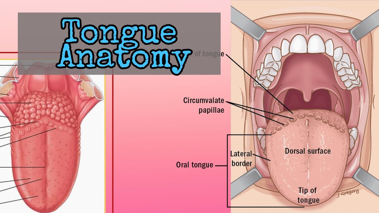
Gross anatomy. The tongue has a tip, dorsum, inferior surface and root. The tongue is made of a midline lingual septum and hyoglossus membrane, and multiple muscles 1,2,4. The muscles are divided into intrinsic and extrinsic muscle groups: The tongue is divided into two parts at the level of the circumvallate papillae 1,3: includes root of.

15.1 Taste Anatomy & Physiology
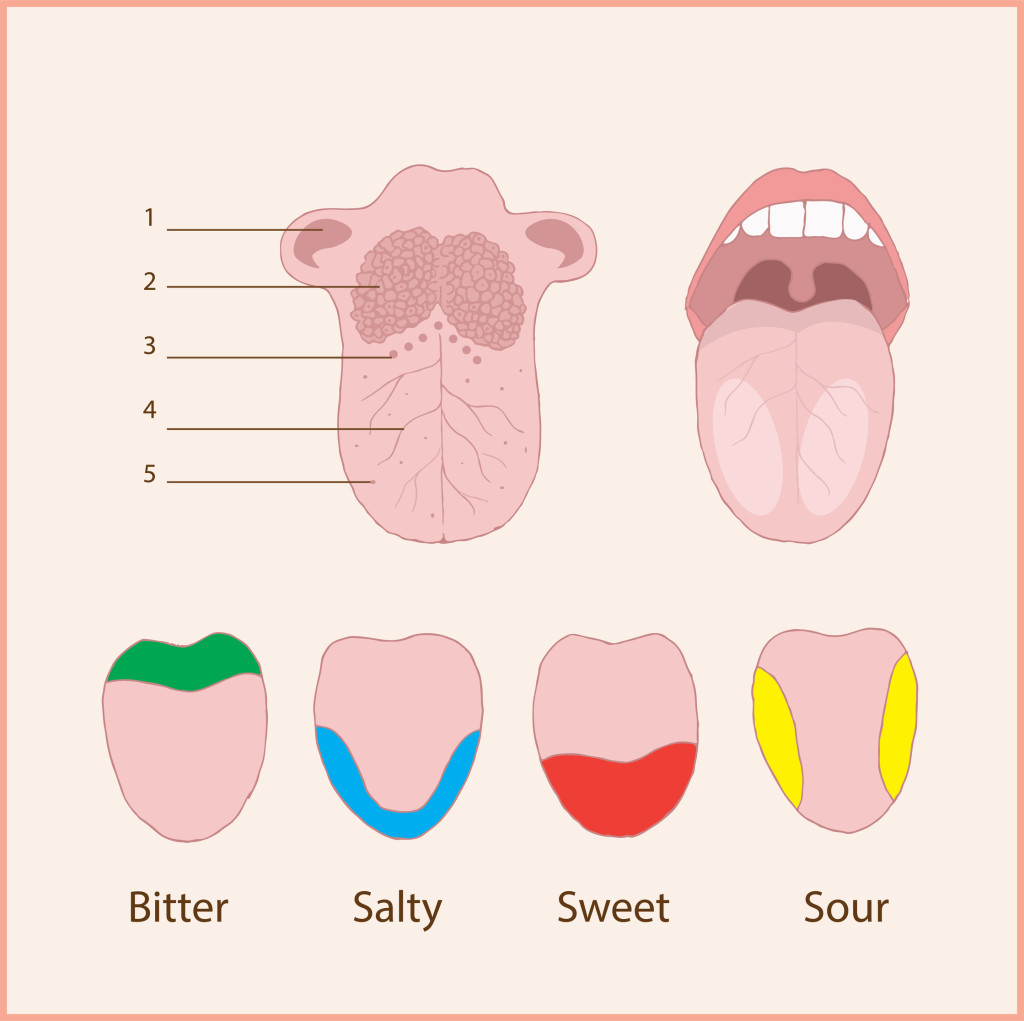
The five areas of the tongue perform different functions and are supported by separate nerves and blood vessels. The five visible parts of the tongue are: Root - The root connects the tongue to the floor of the mouth. It's attached to the hyoid bone and the lower jaw, keeping it in place. Body - The body is the portion of the tongue.
Tongue Anatomy Human with Label coordstudenti
The root of the tongue is posterior and slightly vertical, forming the posterior one third of the tongue. It extends from the hyoid, epiglottis, and soft palate, to the mandible. The body forms the anterior ⅔ of the tongue, and the apex of the tongue is the most anterior end of the body. The entirety of the tongue rests on the mouth's floor.

Tongue Human tongue, Human body anatomy, Anatomy images
The tongue's anatomy includes smooth, moist mucosa on its surface. The surface then contains papillae that provide friction to grasp food particles. Taste buds are located in the papillae, as well.

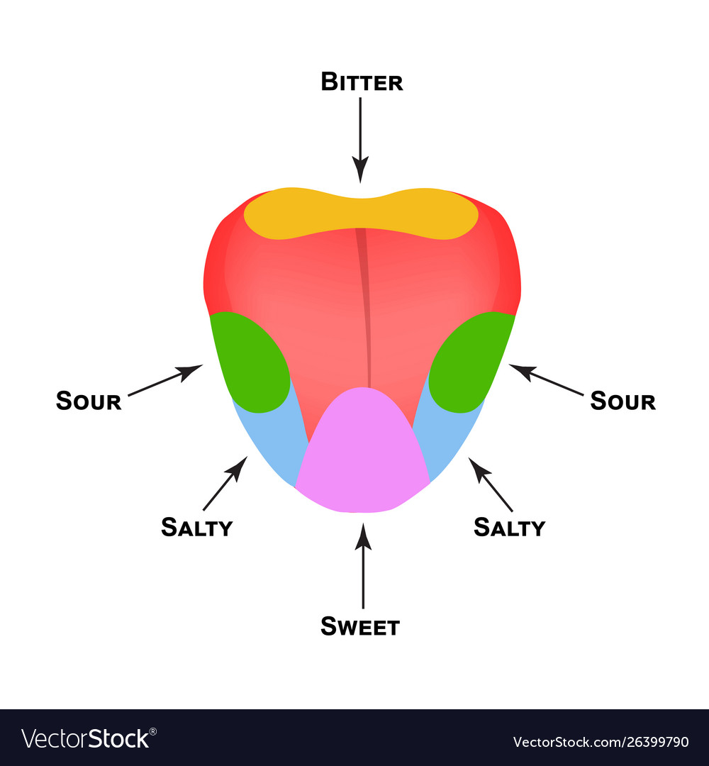
TIL the tongue map that shows parts of the tongue responsible for different aspects of taste is
The tongue (Latin: lingua s. glossa) is a pink-colored, mobile muscular organ of the digestive system. It occupies the oral cavity proper, and from it, the tongue also extends into the oropharynx. The tongue helps to support the floor of the oral cavity, and it helps in food chewing and swallowing and provides tactile, taste and speech.

Human Tongue Anatomy Human tongue, Tongue health, Anatomy
The body (anterior two-thirds of the tongue) is the largest part of the tongue and extends from the sulcus terminalis to the frenulum. The body is further subdivided into anterior and posterior. The anterior body is the area below the hard palate, and the posterior body is underneath the soft palate. A collection of nodular lymphatic tissue.

The Mouth, Pharynx, and Esophagus Anatomy and Physiology II
The human tongue is about 3.3 inches in men and 3.1 inches in women. It is located in the oral cavity. The tongue is divided into three parts: Tip; Body; Base; The tongue is embryologically divided into the anterior and posterior part. The anterior part is known as the oral or presulcal part that includes the root attached to the floor of the.
The Tongue
Therefore, the oral part of the tongue, which arises from the first pharyngeal arch, is innervated by the lingual nerve. Gross anatomy. The tongue is an unpaired structure located within the oral cavity proper. It is bordered: anteriorly and laterally by the upper and lower rows of teeth;
Tongue Surface Anatomy of the Tongue Head and Neck Human Anatomy YouTube
The tongue is a mobile, muscular organ that lies within the mouth and partly extends into the upper throat. The tongue's anatomy is complex; it involves interlacing muscles, nerves, and a blood supply. This article will explain the details of tongue anatomy and how each part contributes to its movements and to functions such as eating, taste.

Tongue Anatomy Human with Label Health Images Reference
The tongue is a muscular organ in the mouth. The tongue is covered with moist, pink tissue called mucosa. Tiny bumps called papillae give the tongue its rough texture. Thousands of taste buds.
In this picture the anterior 2/3 is referred to as the mobile part of the tongue. But what mike
The tongue is a muscular structure in the mouth covered by mucosa whose primary functions are in mastication, taste, and speech. It can be divided into the anterior two-thirds which makes up part of the oral cavity and the posterior-third, part of the oropharynx. 1 The tongue consists of a tip, dorsal surface, ventral surface, and root.
